Pachygyria: The Smooth Brain
Pachygyria is a congenital malformation of the cerebral cortex. It is the result of poor neuronal migration, which causes unusually thick convolutions.
Children with this disease tend to have varying degrees of cognitive impairment, developmental delay, and seizure disorders.
What is the cause, how is it diagnosed and what are the symptoms? Can it be treated? Here’s everything you need to know about pachygyria.
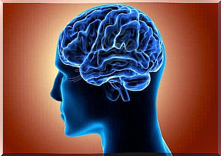
What is the cerebral cortex and what does it look like?
The cerebral cortex covers the surface of the two hemispheres of the brain. It has a structure rich in folds and this allows it to have a wide extension in a small space when compared to the surface of a smooth brain of equal size.
On the surface of the cortex, a series of grooves, fissures or depressions of varying sizes, and convolutions (bulges located between the fissures) can be distinguished.
It consists mainly of glial cells and neuronal bodies ; finally , it is organized in six layers in which the neurons are arranged with extreme precision.
What is pachygyria?
It is a malformation that belongs to the lissencephalic family (disorders in which the surface of the cortex appears smooth). It is located between agiria (total absence of convolutions) and polymicrogyria (malformation of the cortex with too many convolutions).
Pachygyria is a severe congenital malformation (with a very high mortality rate ) for which the organization of the neurons of the cerebral cortex and the structure itself are altered. The cerebral cortex is thinner than normal, since some layers that compose it are absent and the cells inside are poorly organized.
The main feature of this disease is that the cortex has little pronounced cracks and thicker and wider convolutions. This makes its appearance smoother, less wrinkled.
Symptoms
Children with pachygyria usually develop the following symptomatic triad: cognitive impairment, seizures, and severe developmental impairment. They also have a wide variety of symptoms which we will see below.
- Seizures in 90% of children. They usually appear between the fourth and seventh month of life. They are dangerous because they are resistant to any form of treatment.
- Delay and impaired motor and language development. There is usually hypotonia of the legs and arms, which makes walking and standing difficult, and poor speech. Often accompanied by microcephaly.
- Severe to profound cognitive impairment that limits all cognitive functions and autonomy.
- Behavioral and other mental disorders.
How is pachygyria diagnosed?
The absence of characteristic facial or body features can make diagnosis difficult. This, therefore, is based on the clinical examination of the symptoms and on neuroimaging techniques that allow to evaluate the cerebral cortex in detail.
Computed tomography (CT) provides images of the brain in high spatial resolution. However, malformations of the cortex are easier to visualize in vivo. MRI is therefore more useful, as it offers greater contrast and better differentiation of gray matter and white matter regions.
In the images, thickened cerebral convolutions with few superficial fissures are observed, as well as the incomplete development of Silvio’s lateral fissure.
Causes of pachygyria
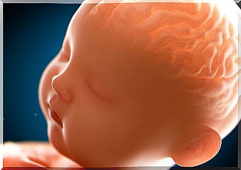
Pachygyria is caused by an interruption of the fetal neuronal migration process. This flaw in the system can be caused by environmental factors or genetic alterations.
Neuronal migration occurs between the seventh and twentieth week of gestation. The aim is the formation of the six layers that make up the cortex. In pachygyria, on the other hand, there are only four layers, which is why the cortex is thinner.
Simplifying, in the brain affected by pachygyria there is the first layer (molecular or plexiform) similar to that of a normal brain. The second, third and fourth layers contain fewer cells than a healthy brain, thus taking on a more subtle structure.
As mentioned, this anomaly in neuronal migration can have environmental causes. These include exposure to certain substances or elements (radiation, alcohol, mercury, etc.).
The genetic alterations responsible for pachygyria are too complex to be addressed here. However, it should be noted that the alterations related to this condition generally affect chromosome 10 and chromosome 17. Similarly, the absence of the LIS1 gene appears to be the cause of lissencephaly.
There is no treatment
As with most rare diseases, there is currently no treatment of the cause, but only aimed at limiting the symptoms. In this case it is essential to take antiepileptic drugs to prevent seizures.
As mentioned, pachygyria is a severe malformation with a fatal outcome; when it does not affect the cortex in its entirety, however, it is compatible with life and the damage will depend on the area affected by the malformation.
In the event of survival, collaboration between multiple specialist figures will be essential. From the physiotherapist who will work on motor alterations such as hypotonia, to the support teacher who will adapt the school program to the child’s abilities.
Naturally, we will have to count on the collaboration of doctors, nurses, occupational therapists and psychologists. The multidisciplinary intervention will allow the child to enjoy a better life by promoting autonomy as much as possible.